


“Nothing in biology makes sense except in the light of evolution”
Theodosius Dobzhansky, 1973
There are upwards of 10 million new cancer cases a year in the world. One in three of us can expect that unwelcome diagnosis, and around one in four will succumb to metastatic, drug resistant disease. The big questions are, why are humans so vulnerable to cancer? what exactly is cancer as a biological process? and why is drug resistance the norm for advanced disease? Evolutionary biology has something to say about each of these grand challenges.
The Hallmarks of Cancer as evolutionary adaptations in a neoplasm[edit]
http://elledgelab.med.harvard.edu/?page_id=305
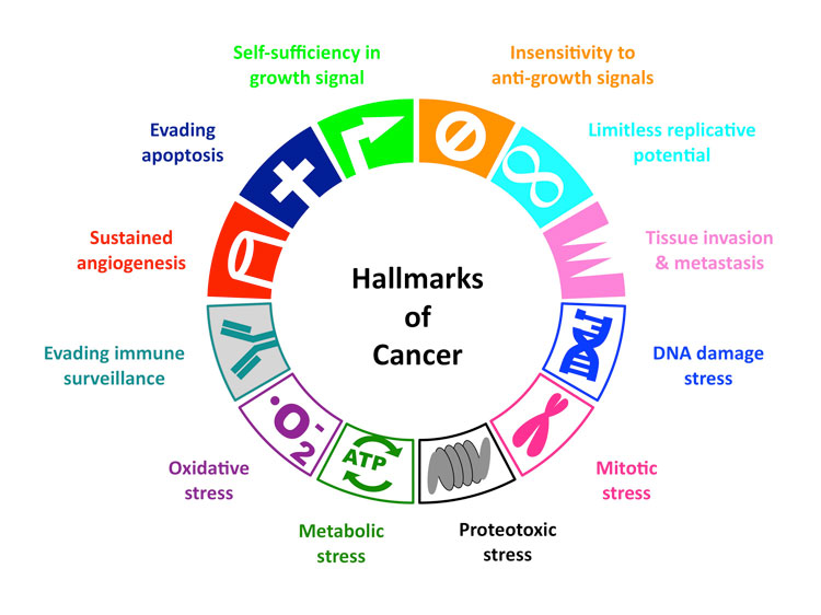
Hallmarks of Cancer
All cancer cells show 6 common traits (10 if you go more in-depth)
They are:
(1) Self-Sufficiency in growth signal, or in other words, they grow and multiply without being told to.
(3) Evading Apoptosis- they refuse to die, like zombies
(4) Limitless Reproduction Potential- They just keep multiplying!
(5) Sustained Angiogenesis- they keep making new blood vessels to feed themselves
(6) Tissue Invasion and Metastasis- they break away from their site of origin and invade surrounding tissues, eventually spreading (metastasizing) throughout the body….like an army of zombies.
Psychological stress can take a tremendous toll on your health. One of the reasons for this is because stress causes inflammation, which in turn is a hallmark of most diseases, from obesity and diabetes to heart disease and cancer.
Six years ago, I interviewed Donald (“Donnie”) Yance, an internationally known herbalist and nutritionist, who shared a really surprising piece of information: stress was actually pinned down as a cause of cancer all the way back in 1908. As Donnie said:
“Eli Jones, the great eclectic physician in cancer, and probably the most brilliant person that ever lived on the face of the planet, wrote a book in 1908 called “Cancer – Its Causes, Symptoms and Treatment.” There isn’t one inaccuracy I can find in that book, written more than 100 years ago.”
In this book, Dr. Jones revealed the top causes of cancer, and the No. 1 cause he listed was unresolved stress. Since then, a number of studies have confirmed this link. In the video above, two doctors at MD Anderson Cancer Center go into some of the details now known about stress and cancer.
Chronic Stress Makes Cancer Spread
Most recently, a study done on mice found that when the animals were chronically stressed, their lymphatic systems underwent changes that allowed cancer to spread more quickly and easily. As reported by Science Alert:1
“Although the study hasn’t been replicated in humans as yet, it’s a huge step towards understanding how stress – which has long been linked to cancer progression – actually helps tumour cells escape…
“Not for a minute are we suggesting that someone who’s just been diagnosed with cancer should not be stressed, because that would have to be one of the most stressful situations”… Erica Sloan from Monash University in Australia, told ABC News.2
“But rather how do we look after cancer patients, because this suggests that stress not only affects patient wellbeing but also gets into the body and affects how the tumour progresses.”
How Does Stress Promote the Spread of Cancer?
Cancer cells typically spread to other areas of the body either via your blood vessels, or through your lymphatic system. Stress hormones affect both of these pathways or channels. Here they were trying to determine how stress hormones affect the spread of cancer cells through the lymphatic system.
The mechanism they found is related to the way adrenaline activates the sympathetic nervous system (SNS) to increase the rate of lymph formation. Adrenaline also causes physical changes in the lymph vessels, allowing cancer cells to migrate into other body parts at a faster rate.
The National Cancer Institute has also previously stated that research with animal models suggests:3
“[Y]our body’s neuroendocrine response (release of hormones into your blood in response to stimulation of your nervous system) can directly alter important processes in cells that help protect against the formation of cancer, such as DNA repair and the regulation of cell growth.”
Other research4,5 has shown that the stress hormone norepinephrine may increase the growth rate of cancer.
Norepinephrine can stimulate tumor cells to produce two compounds (matrix metalloproteinases called MMP-2 and MMP-9) that break down the tissue around the tumor cells, thereby allowing the cells to more easily move into your bloodstream.
Once there, they can travel to other organs and tissues and form additional tumors.
Norepinephrine may also stimulate tumor cells to release a chemical (vascular endothelial growth factor, or VEGF) that aids the growth of the blood vessels that feed cancer cells. This too can increase the growth and spread of the cancer.
Epinephrine — yet another stress hormone — has also been found to cause changes in certain cancer cells, specifically prostate and breast cancer, in ways that makes them resistant to apoptosis (cell death).
How chronic inflammation can lead to cancer
https://www.mdanderson.org/publications/focused-on-health/may-2014/inflamation-cancer-diet.html

August 7, 2015
Chronic inflammation caused by disease or exposure to dangerous chemicals has long been linked to cancer, but exactly how this process takes place has remained unclear.
Timing of inflammation determines whether potentially cancerous mutations may arise.A new study from MIT reveals one reason why people who suffer from chronic inflammatory diseases such as colitis have a higher risk of mutations that cause cancer. The researchers also found that exposure to DNA-damaging chemicals after a bout of inflammation boosts these mutations even more, further increasing cancer risk.
This means that emotional stress can both contribute to the development of cancer and reduce the effectiveness of treatments.
DNA Damage and Oxidative Stress in Human Disease
This special issue contains eight papers, covering several aspects of the implication of DNA damage and oxidative stress. Three papers focus on brain, colorectal, and skin cancers and one paper focuses on the genomic stability and oxidative stress in the cancer-predisposing genetic syndrome ataxia telangiectasia. One paper discusses the implication of nutrients in genomic stability in cell cultures. Two papers discuss the effect of a vitamin and a neuroprotectant on diabetes and traumatic brain injury, respectively. Another paper focused on the impact of autophagy on idiopathic pulmonary fibrosis.
“The vitamin D receptor (VDR) gene polymorphisms in Turkish brain cancer patients” by B. Toptas et al. provides evidence for the first time that the risk of meningiomas might be related to polymorphisms in the nuclear receptor of vitamin D, an important factor for the regulation of cell division and proliferation. In “Oxidative stress in the pathogenesis of colorectal cancer: cause or consequence?” M. Pere reviews the interplay between the several risk factors that have been implicated in colorectal cancer, a very common type of cancer in Western countries, which has a complex etiology. N. C. Jenkins and D. Grossman show, in “Role of melanin in melanocyte dysregulation of reactive oxygen species,” that the presence of melanin in the skin appears to be a double-edged sword: it protects melanocytes as well as neighboring keratinocytes in the skin through its capacity to absorb UV radiation, but its synthesis in melanocytes results in higher levels of intracellular ROS that may increase melanoma susceptibility.
L. B. Ludwig et al. provide that ionizing radiation is more efficient than bleomycin to induce chromosomal instability in ataxia telangiectasia patients and that this instability is not related to a systemic increase in oxidative stress. In “The influence of micronutrients in cell culture: a reflection on viability and genomic stability,” A. L. V. Arigony et al. addresse the effect of several vitamins and minerals by reviewing their role in metabolic routes related to DNA homeostasis. The paper presents lines of evidence whether while in deficiency or excess in cell culture the micronutrients reviewed can reduce or increase the level of DNA damage and influence cell proliferation and viability. Finally, the authors advocate which nutrients should deserve more attention in future studies focusing on the increase of genomic stability and cell fitness under culture conditions.
“Vitamin C intake reduces the cytotoxicity associated with hyperglycemia in prediabetes and type 2 diabetes” by S. I. R. Franke et al. compares the levels of vitamin C intake, which is among the most abundant antioxidants obtained from diet, with the levels of markers of hyperglycemia, DNA damage, and cytotoxicity in subjects with type 2 diabetic or with risk of developing the disease. The authors observe that vitamin C intake slightly higher than the dietary recommendation for healthy individuals can be beneficial to the subjects by preventing the cell death of white blood cells that have been reported in the literature to be associated with diabetes complications.
In “Therapeutic time window for edaravone treatment of traumatic brain injury in mice,” K. Miyamoto et al. deal with the edaravone administration postcontrolled cortical impact (CCT) resulting in a significant reduction in the injury volume and oxidative stress. These findings suggest that edaravone could prove clinically useful to ameliorate the devastating effects of traumatic brain injury (TBI). “Self-eating: friend or foe? The emerging role of autophagy in idiopathic pulmonary fibrosis,” by G. A. Margaritopoulos et al. highlights some key issues regarding the process of autophagy and its possible association with the pathogenesis of idiopathic pulmonary fibrosis.
Advances in molecular biology and bioinformatics are allowing researchers to gain an increased understanding of the function and regulation of genes and to identify pathways that are affected. Currently, the search for biomarkers related to disease is gaining increasing attention and especially biomarkers for oxidative stress and DNA damage became more and more valuable instruments for unraveling disease pathogenesis and facilitating prediction, prevention, and treatment of diseases.
Oncogene-Induced Mitotic Stress: p53 and pRb Get Mad Too
Proteotoxic stress of cancer: Implication of the heat-shock response in oncogenesis
Article
Heat-mediated reduction of apoptosis in UVB-damaged keratinocytes in vitro and in human skin ex vivo
- “Heat stress can also trigger extensive denaturation, degradation and aggregation of critical intracellular proteins [15, 16], leading to defective DNA replication, transcription and repair, thus affecting cell survival and apoptosis [17][18][19]. The deleterious effect of heat on cellular processes is normally countered by activation of a conserved heat shock response [20][21][22], which stabilises cells by interacting with pro-survival signalling pathways such as PI3K/Akt [20, 23, 24]. Continuous exposure to heat stress independently or concurrently with UV radiation is commonly experienced in several geographical locations. “
‘Master switch’ helps cancer cells survive stress
New survival mechanism could be a target for future cancer drugs
- Date:
- December 3, 2015
- Source:
- Institute of Cancer Research
- Summary:
-
Scientists have discovered a ‘master switch’ within cancer cells that seems to override the normal stress response and allows them to survive conditions that would normally be lethal. The mechanism could be critical in allowing cancer cells to withstand the huge amounts of stress they come under as they divide rapidly and their metabolism goes into overdrive.
Scientists have discovered a ‘master switch’ within cancer cells that seems to override the normal stress response and allows them to survive conditions that would normally be lethal.
Psychological stress can take a tremendous toll on your health. One of the reasons for this is because stress causes inflammation, which in turn is a hallmark of most diseases, from obesity and diabetes to heart disease and cancer.
Six years ago, I interviewed Donald (“Donnie”) Yance, an internationally known herbalist and nutritionist, who shared a really surprising piece of information: stress was actually pinned down as a cause of cancer all the way back in 1908. As Donnie said:
“Eli Jones, the great eclectic physician in cancer, and probably the most brilliant person that ever lived on the face of the planet, wrote a book in 1908 called “Cancer – Its Causes, Symptoms and Treatment.” There isn’t one inaccuracy I can find in that book, written more than 100 years ago.”
In this book, Dr. Jones revealed the top causes of cancer, and the No. 1 cause he listed was unresolved stress. Since then, a number of studies have confirmed this link. In the video above, two doctors at MD Anderson Cancer Center go into some of the details now known about stress and cancer.
Chronic Stress Makes Cancer Spread
Most recently, a study done on mice found that when the animals were chronically stressed, their lymphatic systems underwent changes that allowed cancer to spread more quickly and easily. As reported by Science Alert:1
“Although the study hasn’t been replicated in humans as yet, it’s a huge step towards understanding how stress – which has long been linked to cancer progression – actually helps tumour cells escape…
“Not for a minute are we suggesting that someone who’s just been diagnosed with cancer should not be stressed, because that would have to be one of the most stressful situations”… Erica Sloan from Monash University in Australia, told ABC News.2
“But rather how do we look after cancer patients, because this suggests that stress not only affects patient wellbeing but also gets into the body and affects how the tumour progresses.”
How Does Stress Promote the Spread of Cancer?
Cancer cells typically spread to other areas of the body either via your blood vessels, or through your lymphatic system. Stress hormones affect both of these pathways or channels. Here they were trying to determine how stress hormones affect the spread of cancer cells through the lymphatic system.
The mechanism they found is related to the way adrenaline activates the sympathetic nervous system (SNS) to increase the rate of lymph formation. Adrenaline also causes physical changes in the lymph vessels, allowing cancer cells to migrate into other body parts at a faster rate.
The National Cancer Institute has also previously stated that research with animal models suggests:3
“[Y]our body’s neuroendocrine response (release of hormones into your blood in response to stimulation of your nervous system) can directly alter important processes in cells that help protect against the formation of cancer, such as DNA repair and the regulation of cell growth.”
Other research4,5 has shown that the stress hormone norepinephrine may increase the growth rate of cancer.
Norepinephrine can stimulate tumor cells to produce two compounds (matrix metalloproteinases called MMP-2 and MMP-9) that break down the tissue around the tumor cells, thereby allowing the cells to more easily move into your bloodstream.
Once there, they can travel to other organs and tissues and form additional tumors.
Norepinephrine may also stimulate tumor cells to release a chemical (vascular endothelial growth factor, or VEGF) that aids the growth of the blood vessels that feed cancer cells. This too can increase the growth and spread of the cancer.
Epinephrine — yet another stress hormone — has also been found to cause changes in certain cancer cells, specifically prostate and breast cancer, in ways that makes them resistant to apoptosis (cell death).
This means that emotional stress can both contribute to the development of cancer and reduce the effectiveness of treatments.
Center, linked below, “copper-binding molecules [ceruloplasmin, heparin, and tripeptide glycly-histadyl-lysine] are non-angiogenic when free of copper, but they become angiogenic when bound to copper.”
On January 21, 2000, the University of Michigan reported that researchers had “successfully stopped the growth and spread of cancer by depriving the tumors of the copper supply they need to form new blood vessels.” Dr. George Brewer used an inexpensive compound called tetrathiomolybdate (TM) to lower the serum copper levels in patients with cancer. This study was done with a group of 18 patients in hospice with 11 different types of metastatic cancer. The goal of the study was to reduce ceruloplasmin to 20% of baseline for at least 90 days. The treatment achieved this goal in 6 patients, and 5 of those patients have seen no tumor growth or new tumors for more than 2 years. The other 12 patients could not achieve the target copper levels, suggesting that it can take more than a month to reduce copper levels to target, during which time the cancer may progress rapidly.
A larger 100 patient Phase II trial of TM is currently underway. “This is not a cure for cancer, but a disease stabilizer.” Dr. Brewer said of the anti-copper drug. Dr. Brewer sought and received orphan drug status by the FDA for AmmoniumTetrathiomolybdate on January 31, 1994 for the treatment of Wilson’s disease.
Based on the research in Michigan, Dr. Brewer said “There’s no reason to try to follow a low-copper diet. The only two foods we ask people not to eat are liver, which is loaded with copper and shellfish, which have intermediately high amounts.” The research does not suggest that the copper in most food and supplements help promote cancer. “You’re not trying to get rid of all the copper, people die. [What you’re doing with zinc or TM therapy is] reducing the excessive load of copper and preventing it from reaccumulating.”
Our recommended compounding pharmacist for the TM:
Wayne Loveland, pharmacist
The Prescription Center
1907 West Avenue South
LaCrosse, WI 54601
608-788-4500
800-203-9066
608-788-4501 fax
Click on links below to learn more.
Quick Study-
“Copper-lowering drug stabilizes advanced cancer in anti-angiogenesis trial”
This is a news release dated 1/20/2000 from the University of Michigan regarding copper, cancer and tetrathiomolybdate (TM).
“Copper and Cancer”
This is from an article in the Journal of the American Medical Association dated 2/23/2000, regarding copper and cancer.
For more in-depth information see below-
“Treatment of Metastatic Cancer with Tetrathiomolybdate, an Anticopper, Antiangiogenic Agent: Phase I Study”
This is a research paper dated January 2000, authored by George J. Brewer, M.D. and R. D. Dick, et al., regarding copper and TM. Dr. Brewer, a University of Michigan human genetics professor and researcher, originated the work on the use of TM and cancer. Abstract is also available.
“Angiogenesis and Cancer Control: From Concept to Therapeutic Trial”
This is a research paper published in 1999, authored by Steven Brem, M.D., of the H. Lee Moffitt Cancer Center & Research Institute. See Table 2 (showing copper as a trace element), Table 7 and the discussion concerning copper under the subtitle “Copper Antagonists/Chelators.”
Estrogen dominance is a term that describes a condition where a woman can have deficient, normal or excessive estrogen, but has little or no progesterone to balance its effects in the body. Even a woman with low estrogen levels can have estrogen dominance symptoms if she doesn’t have enough progesterone.
Excess estrogen causing estrogen dominance is also received transdermally from all sorts of external sources. These are called Xenoestrogens. These are fat-soluble and non-biodegradable in nature. The major sources of these Xenoestrogens are pesticides, detergents, petroleum products, plastic products, cosmetics, even spermicides used for birth control in diaphragm jellies, condoms and in vaginal gels. So think twice when you drink your hot coffee or tea in that plastic or Styrofoam cup from the convenient store on your way to work. What ever you do, DO NOT heat your food in plastic cookware. All of these contribute to the estrogen dominance and have been linked to birth defects in both humans and animals.
Estrogen Dominance and the symptoms associated with it:
- Acceleration of the aging process
- Allergies, including asthma, hives, rashes, sinus congestion
- Autoimmune disorders such as lupus erythematosis, thyroiditis,and Sjoegren’s
- Breast cancer
- Breast tenderness
- Cervical dysplasia
- Cold hands and feet as a symptom of thyroid dysfunction
- Copper excess
- Decreased sex drive
- Depression with anxiety or agitation
- Dry eyes
- Early onset of menstruation
- Endometrial (uterine) cancer
- Fat gain, especially around the abdomen, hips and thighs
- Fatigue
- Fibrocystic breasts
- Foggy thinking
- Gallbladder disease
- Hair Loss
- Headaches
- Hypoglycemia
- Increased blood clotting (increasing risk of strokes)
- Infertility
- Irregular menstrual periods
- Irritability
- Insomnia
- Magnesium deficiency
- Memory loss
- Mood swings
- Osteoporosis
- Polycystic ovaries
- Premenopausal bone loss
- PMS
- Sluggish metabolism
- Thyroid dysfunction mimicking hypothyroidism
- Uterine cancer
- Uterine fibroids
- Water retention & bloating
- Zinc deficiency
When Estrogen Dominance occurs within the body these are some of the results:
- Endometriosis
- Blood Clots
- Elevated Blood Pressure
- Fibroid Breasts
- Infertility
- Irregular Menstrual Flow
- Uterine Fibroids
- Breast Tenderness
- Mood Swings
- Uterine Cancer
- Hair Loss
- Depression
- Weight Gain
- Migraine Headaches
- Spotting
- Breast Cancer Risk
- Insomnia
- Inflammation
- Abnormal Pap Smears
- Fluid Retention
- Cramping
- Vaginal Dryness
- Thyroid Imbalances
- Decrease in Memory
- Low or No Sex Drive
Estrogen dominance symptoms can be greatly improved and even be reversed by balancing the excess estrogen and reversing the estrogen dominance with natural bioidentical progesterone. One of the best sources of Natural Bioidentical Progesterone is Wild Yam (Dioscorea Villosa). Wild Yam contains a compound Diosgenin which is the active biochemical constituent with progesterone properties. This herb has been used for years as the base for synthetic hormonal drugs which when put through the chemical process create more harmful side effects when taken. However, in its natural unadulterated form, Wild Yam can work wonders when it is converted into bio identical progeterone.
But if it’s so wonderful then why don’t you hear more about it? The answer is easy! The pharmaceutical companies can not make big bucks on an herb that is chemical free. No chemical processes then no patent. When pharmaceutical companies can patent a drug it becomes exclussive with a price that they set. The pharmaceutical companies would rather process products that create nasty side effects, fund studies that support these products then they sell another drug to combat the side effects of the first and so on. The results: A damaging viscous downward cycle of health.
We are all–men, women and children–suffering from Estrogen Dominance, because there is so much of it in our environment. You would have to virtually live in a bubble to escape the excess estrogens we’re exposed to through pesticides, plastics, industrial waste products, car exhaust,meat, soaps and much of the carpeting, furniture and paneling that we live with indoors every day. You may have on-and-off sinus problems, headaches, dry eyes, asthma or cold hands and feet for example, and not know to attribute them to your exposure to Xenohormones. Over time the exposure will cause more chronic problems such as arthritis and premature menopause symptoms, and may be a direct or indirect cause of cancer.
Estrogen dominance is when your body is subjected to excess estrogen like compounds usually from a source outside the body. Estrogen dominance can be reduced by supplementing with Bioidentical progesterone cream and by reducing your exposure to environmental toxins or xenohormones.
Natural Alternative for Estrogen Dominance
Bioidentical progesterone cream (Progensa 20)
Recent studies indicate that estrogen dominance is reversed by bioidentical progesterone cream by curbing hormonal imbalances, which are caused by exposure to xenohormones(external sources of estrogen)and your own body when your liver is not functioning properly and it allows estrogen levels to build up and cause estrogen dominance.
ProSoothe is an all natural herbal formula that significantly improves estrogen dominance, uterine fibroids and pelvic pain/cramps, irritability, tension, mood swings, acne, headaches, breast pain, bloating and weight gain.
Also found in this synergistic herbal formula is dandelion and vitex,(chaste tree) that helps the body remove exogenous,(external excess estrogen)/estrogen dominance from hormone therapy or contaminated food,(xenosteroids).
Beyond the treatment of liver disorders, everyday care of the liver lays a cornerstone for total body health. Naturopaths and others who look beneath the symptoms of an illness to its underlying cause, often discover that the liver has had a role to play. This is true across a vast range of different ailments including estrogen dominance.
Through my research I have also learned, watermelon has a great amount of glutathione. Glutathione can protect us from cancer. It is an antioxidant. While other fruits, that also have glutathione and are rich in antioxidants are berries, oranges, pomegranate, apricots, prunes, avocado, grapefruit, strawberries and peaches. Cinnamon, asparagus, legumes, nuts, spinach and bell peppers can also make our skin healthier. We often hear glutathione when we talk about skin lightening products.
What we don’t know is we can eat cystine-rich food because our body uses cystine and converts it to glutathione. Cystine-rich food includes dairy products such as cheese, yogurt and chicken breast. With cystine-rich food and foods rich in selenium, the production of glutathione in our body is increased. Tuna, beef, chicken, turkey, eggs, cheese, and Brazil nuts are good source of selenium. Brazil nuts are especially rich in selenium, which helps increase the body’s production of glutathione. Other foods rich in selenium include tuna, beef, walnuts, eggs, cottage cheese, and turkey.
To make our skin more beautiful, we can eat food with B-complex. Watermelons also contain Alkaline Water which is all the rage these days. Alkaline water removes toxins and acidity from the body. Some say it also reduces food cravings that leads to weight loss.
Aside from glutathione and alkaline water, watermelon is also rich in beta carotene, potassium, vitamin A, and vitamin B. Personally, I am surpised at how much we can get from watermelons as I reckon it is only made up of water. From now on, next to bananas – let’s add watermelons in our fruits baskets!
GENE-NUTRIENT AND NUTRIENT-NUTRIENT INTERACTIONS
Genetic analyses can provide further insight to assessing nutrient-cancer relations. Studies of folate and colorectal cancer provide a useful illustration. A common polymorphism in the gene that codes for the enzyme methylenetetrahydrofolate reductase (MTHFR) leads to a variant enzyme with impaired ability to convert 5,10-methylenetetrahydrofolate to 5-methylenetetrahydrofolate. Our group and others found a strong link between this polymorphism and risk of colorectal cancer (7). Men who are homozygous for the variant had about one-half the risk of colorectal cancer as did other men. In further analyses, we found that this association was limited to those without folate deficiency in whom the homozygous variant carried about a two-thirds reduction in risk. These findings provide powerful support for a causal interpretation of the link between folate and colorectal cancer. The presence or absence of the variant gene is almost certainly unrelated to dietary intake of folate or any other personal characteristic. Thus, it is difficult to attribute any observed associations of that gene with colon cancer risk to confounding factors. A further finding of a biologically plausible interaction between this variant enzyme and folate status renders a noncausal interpretation implausible. Without evidence from this gene-nutrient interaction, one might plausibly interpret the relation of high folate status to lower risk of colorectal cancer to other healthy behaviors that are correlated with folate intake. The finding of gene-nutrient interactions substantially reduces these concerns.
Another example illustrating a different aspect of gene-nutrient interactions is the relation between antioxidants and risk of prostate cancer. Previous observational studies showed that men with low circulating concentrations of vitamin E, lycopene, and selenium have a higher risk of future development of advanced prostate cancer (8, 9). All 3 of these nutrients are potent antioxidants. Vitamin E and selenium were shown in randomized trials to reduce the risk of prostate cancer (10, 11). However, these trial results have not been fully accepted, largely because neither trial specified prostate cancer reduction as a primary a priori outcome. Both of these agents are currently being tested in a large randomized trial, the Selenium and Vitamin E Cancer Prevention Trial (SELECT) (12). A common polymorphism with apparently functional consequences has been identified in the gene coding for manganese superoxide dismutase, the principal antioxidant enzyme in mitochondria. Because of the strong hypothesis that antioxidants might be important for prostate cancer risk, our group studied the effect of this polymorphism. Overall, we found no significant association. However, we found a highly statistically significant interaction between the variant genotype and these 3 antioxidants, such that men homozygous for the variant allele were remarkably sensitive to dietary antioxidants (13). We observed a 10-fold gradient in risk comparing those in the highest quintile of a combined antioxidant score (based on blood concentrations of vitamin E, lycopene, and selenium) with those in the lowest quintile, but only among men homozygous for the variant allele. This kind of gene-nutrient interaction, analogous to folate-MTHFR, strongly supports a causal interpretation for these antioxidants. However, of note, no main effect was observed for the variant allele. Instead, we found that the variant defined a subgroup that is quite sensitive to exogenous antioxidants. This finding suggests that SELECT may fail to observe an overall effect if most of the benefit is limited to the minority of men who are homozygous for the variant allele. Unlike in studies of drugs, which are novel exogenous agents, one might strongly expect the presence of gene-nutrient interactions because the nutrients have long been part of human evolutionary history. Because of the issues of timing and duration described earlier, gene-nutrient interactions may potentially provide sufficiently strong evidence for dietary recommendations if definitive randomized trials are infeasible.
Nutrient-nutrient interactions are also likely. For example, in the Calcium Polyp Prevention Trial, Grau et al (14) observed a significant reduction in polyp recurrence in the calcium group compared with the placebo group, but the effect was limited to those with 25-hydroxyvitamin D values above median [29.1 ng/mL; relative risk = 0.71; 95% CI: 0.57, 0.89] and not seen in those with lower 25-hydroxyvitamin D concentrations [relative risk = 1.05; 95% CI: 0.85,1.09]. Thus the nutrient status of the trial participants may affect the results of an intervention trial using a different nutrient in unpredictable ways.
David Ewens discusses the causes of mineral deficiencies and details the most effective & Bio-available (absorbable) minerals to use as the basis for health.
He also covers the little-known fact that rock derived Elemental minerals/metals (iron,zinc, etc) that are commonly used in supplements & mineral water are actually dangerous to the body.
There are approx 72 Minerals that are essential for fundamental health processes. Without them, the bodily functions can start to go out of balance.
All dis-ease begins with mineral deficiency.
These particular mineral bases from Neyharting moor are the best products i have come across. Plus the Neways International product called Feroxin See Video:
Preview YouTube video Mineral Deficiencies – Causes & Effects


Tumor Cellular Escape from Immune Surveillance – YouTube
Zaira Kadaghidze (Moscow) Conference in remembrance of prof. Anatoliy Baryshnikov (21.09.1944–31.05.2015 …
Survival of the sneakiest: how tumours escape immune attack …
6.5 – Cancer: Evading the immune system – YouTube
https://www.youtube.com/watch?
|
The female hormone estrogen seems to play a key role. Women with high estrogen levels in their blood have increased risk for breast cancer. Since exercise lowers blood estrogen, it helps lower a woman’s breast-cancer risk. Exercise also reduces other cancer-growth factors such as insulin. https://www.fredhutch.org/en/events/healthy-living/Trim-Risk.html
|

“Notice the feeling of being at a turning point in the nature of our species. Upheavals around you in the economy and elsewhere are symptoms of far deeper evolu…tion. Visualize shifting from Homo sapiens to Homo luminous, that is, at a cellular level, emerging as beings with ability to perceive vibration and light that creates physical worlds at a much higher level. See the grid or holodeck of energy that reveals what is behind what you think. You are a wizard projecting from behind your own curtain. Answers to longstanding mysteries of existence are arising like the phoenix within. Evolution is accelerating not between generations, but within a generation. This shatters beliefs about how evolution works, inviting you to question all beliefs and shift attention to deeper, innate knowing. Imagine a biological and spiritual quantum leap in this life. The caterpillar does not know what is coming. Trust in the process.” – Liara Covert
|
This film is good except for one part that the narrator had it all wrong. Starting at nine 9 minutes and 15 seconds into the video, she said that “individual” plant protein do not contain all the essential and none essential amino acids that animal protein has. This is wrong and even dietitians also get it wrong. The truth is that all individual plants contains all the essential and non-essential amino acids in just like animal proteins.
This fact is very important because if people don’t know the truth, the meat industry your ignorant doctor can always use this lie to tell vegans that individual plants don’t have all the proteins you need. This is a lie and unfortunately even vegans like this narrator still believe in this kind of lies spread by dietitians and doctors. Check out Dr John McDougall’s article in which he addressed this lie full on: http://www.drmcdougall.com/
Preview YouTube video a delicate balance – the truth..
cate balance – the truth..
|

Jan 21, 2017 – Uploaded by High Intensity Health27:33 Pro-Oxidant Therapies: Oxidative stress is a double edged sword for cancer. Cancer cells at a basal …Nutrition and Metabolic Stress – YouTube
Apr 19, 2016 – Uploaded by Geetesh Vemurimetabolic stress workout metabolic stress cancer metabolic stress and trauma open abdomen metabolic stress …
Preview YouTube video The top 10 cancer causing food ingredients to AVOID

Cancer-Causing Substances in the Environment

Cancer is caused by changes to certain genes that alter the way our cells function. Some of these genetic changes occur naturally when DNA is replicated during the process of cell division. But others are the result of environmental exposures that damage DNA. These exposures may include substances, such as the chemicals in tobacco smoke, or radiation, such as ultraviolet rays from the sun.
People can avoid some cancer-causing exposures, such as tobacco smoke and the sun’s rays. But others are harder to avoid, especially if they are in the air we breathe, the water we drink, the food we eat, or the materials we use to do our jobs. Scientists are studying which exposures may cause or contribute to the development of cancer. Understanding which exposures are harmful, and where they are found, may help people to avoid them.
The substances listed below are among the most likely carcinogens to affect human health. Simply because a substance has been designated as a carcinogen, however, does not mean that the substance will necessarily cause cancer. Many factors influence whether a person exposed to a carcinogen will develop cancer, including the amount and duration of the exposure and the individual’s genetic background. Learn more about Environmental Carcinogens and Cancer Risk.
- Aflatoxins
- Aristolochic Acids
- Arsenic
- Asbestos
- Benzene
- Benzidine
- Beryllium
- 1,3-Butadiene
- Cadmium
- Coal Tar and Coal-Tar Pitch
- Coke-Oven Emissions
- Crystalline Silica (respirable size)
- Erionite
- Ethylene Oxide
- Formaldehyde
- Hexavalent Chromium Compounds
- Indoor Emissions from the Household Combustion of Coal
- Mineral Oils: Untreated and Mildly Treated
- Nickel Compounds
- Radon
- Secondhand Tobacco Smoke (Environmental Tobacco Smoke)
- Soot
- Strong Inorganic Acid Mists Containing Sulfuric Acid
- Thorium
- Vinyl Chloride
- Wood Dust
